Application of terahertz imaging and spectroscopy in medical field
terahertz imaging; terahertz spectroscopy; biomedicine; traditional Chinese medicine
Abstract
In the medical field, the research on terahertz imaging and spectroscopy is no longer the basic research of a few years ago, but the intensive research on tissue, cell, skin imaging and TCM detection and analysis.
Compared with traditional medical means, such as ultrasound, X-ray, NUCLEAR magnetic resonance and infrared ray, Terahertz wave technology has more significant advantages, mainly reflected in strong broadband spectral analysis ability, high resolution and low energy consumption. In this paper, the application of Terahertz technology in biomedicine and traditional Chinese medicine is reviewed, and the status and development of terahertz technology in human skin tissues, tumors, cancers and Chinese herbs are analyzed. Based on the above research progress, terahertz imaging technology has laid a foundation for its application in the medical field.
0 Introduction
Terahertz wave (terahertz, THz) refers to electromagnetic waves with a frequency of 0.1-10 THz, a wavelength of 0.03~3 mm, and a wave number of 3.33-333 cm-1. This wave band is between millimeter waves and infrared.
Terahertz technology combines the characteristics of the two fields of photons and electronics, which covers different disciplines such as material engineering, optics, chemistry and physics, and is an interdisciplinary discipline.
In recent years, with the continuous in-depth research of domestic and foreign scholars on terahertz technology, this technology has been widely used in communications and military affairs, and at the same time, it has also achieved more results in the medical field. According to the characteristics of signal processing and detection principles, terahertz technology can be divided into terahertz spectroscopy and terahertz imaging technology. These two technologies are not only used for nuclear magnetic resonance imaging, but also for skin cancer detection.
Compared with X-rays, terahertz waves have lower photon energy (1 THz=4.1 meV), which can avoid the side effects of X-ray medical imaging.
In addition, water molecules show strong absorption for terahertz waves. Therefore, in medicine, terahertz spectroscopy technology can be used to observe the difference between the water content in tumor tissue and normal cells, so as to judge the development of tumors. The amount of water content differentiates between normal tissue and inflamed skin tissue.
This paper sorts out the terahertz time-domain spectroscopy technology and terahertz imaging technology, summarizes the research status of terahertz in epidermal tissue, tumor research, biological cells, and traditional Chinese medicine, and makes a summary of its development direction in the medical field outlook.
1 Terahertz Spectroscopy and Imaging Technology
1.1 Terahertz time-domain spectroscopy technology
Terahertz time domain spectroscopy (THz-TDS) technology is a new spectral detection method, which uses ultrashort pulses of transmitted or reflected terahertz pulses in the time domain to measure the temporal changes of the generated electromagnetic field.
The basic system of this technology consists of a time delay control system, an emitting crystal, a terahertz detection device, a beam splitter and a femtosecond laser. Figure 1 is a schematic diagram of the basic structure of the THz-TDs system.
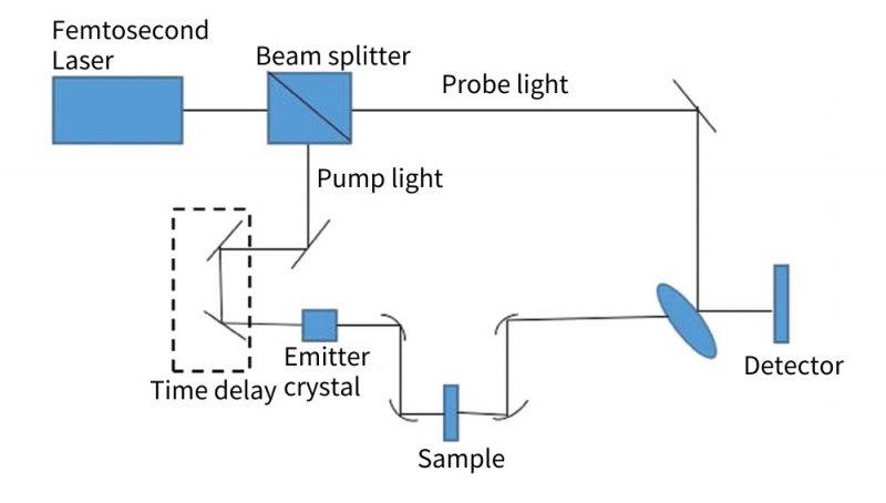
Fig. 1 Terahertz time domain spectroscopy system
During the working process, the pulse is sent out by the femtosecond laser, and then it becomes two paths under the action of the beam splitter, that is, the detection pulse and the pump pulse. Among them, the pump pulse will pass through the emitting crystal under the action of the time delay system emit terahertz pulses.
The detection pulse will pass through the terahertz detector, and then drive the terahertz detection to complete the detection of the pulse intensity. By adjusting the time delay coefficient, the delay between the above two pulses can be changed, and then the terahertz pulse in the frequency domain waveform and the overall time domain can be obtained.
After a certain Fourier transform, the frequency domain spectrogram of the sample to be tested can be drawn. Based on this, various specific optical parameters such as projection, refraction and absorption are obtained.
Based on the use of different detection methods for different samples, THz time-domain spectroscopy systems can be divided into two categories: “transmissive” and reflective systems. The schematic diagram of the terahertz time-domain system is shown in Figure 2.

Fig.2 Schematic diagram of terahertz time domain system
These two types of systems have almost the same working principle. Under the action of the beam splitter, the emitted femtosecond laser pulses are divided into two paths:
- Pumping light, whose function is to emit ultrashort wave pulses;
- The detection light is used to detect the instantaneous electric field amplitude generated by the terahertz pulse in the frequency domain and time domain.
By changing the delay time between the scanning laser and the terahertz pulse, the waveform of the energy intensity in the pulsed electric field at different times is obtained.
The schematic diagram of the transmission terahertz system is shown in Figure 2a. This system is widely used in the research of materials, including electrolytes, conductors, superconductors, organic and inorganic materials, liquefied gases, and flames. The difference between the two is that the detector in the reflective system mainly receives the terahertz waves reflected from the sample surface. Figure 2b is a schematic diagram of the principle of the reflective terahertz time-domain system. Based on this principle, in practical applications, the required spectroscopic system is selected according to different needs.
In the medical field, researchers use terahertz spectroscopy to obtain the optical properties (absorption and refraction parameters) of samples in the terahertz band. The study of terahertz waves in the vibration (rotation-vibration) transition of macromolecules can also be used to obtain biological information at the molecular level.
At present, THz-TDS, as a new technology, has been widely used in the fields of biomedicine and Chinese herbal medicine. In biomedicine, it can measure the characteristics of different biological tissues, analyze chromosomes, cells and proteins, and analyze and detect the genetics of organisms; in Chinese herbal medicine, it can judge the different absorption degrees of drugs for spectra.
1.2 Terahertz imaging technology
The main working principle of terahertz imaging technology is to irradiate the sample with terahertz waves, analyze and process the reflection and transmission information generated by it, and then draw terahertz images. The basic imaging system consists of computer system, data collector, terahertz detection device, optical system and light source. According to different waveforms, terahertz imaging can be divided into two types: pulse imaging and continuous wave imaging.
Among them, the working principle of pulse imaging and time-domain spectroscopy is similar, that is, the detection element converts the signal containing position information into an electrical signal. And convert the image processing unit into a two-dimensional image 4 by the electric signal, so as to obtain the distribution of the internal refractive index of the object. The continuous wave is aimed at the edge damage of the sample or the internal structure of the sample. After the scattering of the terahertz wave, the intensity of the electromagnetic field has a different distribution. The damage location, internal material and shape are judged, and the system complexity and cost are low.
When imaging objects, in order to improve the stability and accuracy of measuring terahertz pulses, the researchers combined differential imaging technology with it. In 2007, B. Pradarutti et al. determined the development plan of the differential imaging system as shown in Figure 3, and the proposed optical path system is shown in Figure 3a.

Fig. 3 Terahertz differential imaging system
The differential imaging is completed by 2 groups of 8 InGaAs photodiodes to form a line-mounted optical probe balance detection device with 8 pixels. Figure 3b is a schematic diagram of the 8-channel balance detection device at that time. The pixels generated by each photodiode in the probe have a diameter of 0.5 mm, and the two adjacent pixels have a distance of 0.75 mm. The corresponding pixel photoelectric signal intensity output from the linear photodiode array on both sides of the detector is subtracted. Then the differential signal strength D6-4 corresponding to 8 pixels can be obtained.
In the continuous research on terahertz pulse imaging, it is found that the imaging quality will be affected by polarization factors to a large extent. The imaging scheme formulated according to the polarization state of the terahertz electric field is shown in Figure 4.
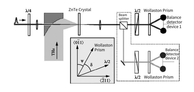
Fig. 4 Terahertz polarization system
Through the <111> crystal plane The detection of ZnTe crystal is carried out by cutting. The detection light first passes through a quarter of the wave plate, and then passes through the small hole on the off-axis parabolic mirror perpendicular to the horizontal line and enters the ZnTe crystal together with the terahertz light wave, and then exits. The probe light continues to pass through the half-wave plate and the Wollaston prism (Wollaston prism, WP), and finally enters the balance detection device.
By adjusting the angle between the wave plate and WP, the polarization intensity generated by the terahertz electric field can be changed. In addition, it can also be seen from the experiment that the light emitted by the non-polarized beam splitter detects another polarized beam.
With the gradual maturity of terahertz imaging technology, people have higher requirements for image resolution.
Researchers soon worked on self-mixing coherent imaging and confocal imaging. The former is to judge the imaging depth of the sample and can be used to observe the incision size of the wound. The latter is to increase the resolution of the samples. The frequency mixing imaging system and the confocal imaging system are shown in Figure 5 and Figure 6.
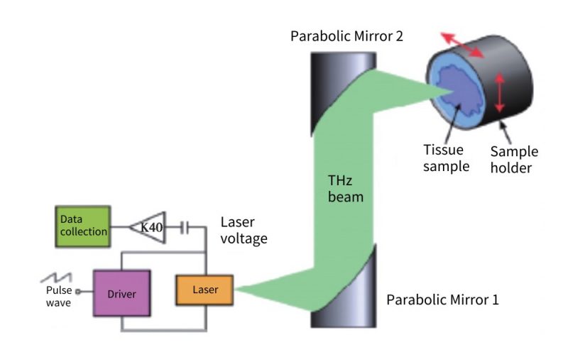
Fig. 5 Mixing imaging system
Fig. 6 Confocal imaging system
2 Applications of terahertz imaging and spectroscopy in the medical field
THz wave has low photon energy. The photon energy of 1THz is about 4meV, and it will not cause tissue damage to biological ionization. Therefore, non-destructive imaging of substances can be realized, and small wounds and non-ionization safety detection of human body can be realized. It has broad application prospects in biomedicine.
With the further development of THz-TDS technology in the field of imaging, the quality, stability, resolution and depth of its images are gradually improving.
At present, terahertz imaging research focuses on the lesion properties of tumor tissues, such as skin basal cell carcinoma, liver cancer, cervical cancer and other cancerous cells. The United States, Australia, and China have achieved remarkable results in this regard.
Compared with traditional imaging technology, terahertz imaging technology has higher spatial resolution and penetration, which can be used for identification and composition analysis of opaque materials, and can realize non-destructive testing of samples at the same time. Therefore, terahertz has great application value and development potential in medical detection and diagnosis.
2.1 Application of terahertz spectroscopy
2.1.1 Amino acids and peptides
The terahertz band is directly related to the energy absorption of amino acids and polypeptides.
According to the particularity of the terahertz band, the energy absorption stage of amino acids and polypeptides can be directly judged.
With the current development of terahertz technology, it has achieved good results in the research of amino acids and peptides. Liu et al. used the THz-THS system to collect different phase spectra of different amino acids, including alanine, methionine, leucine and valine, distributed at 0.1-3 THz, as shown in Figure 7.
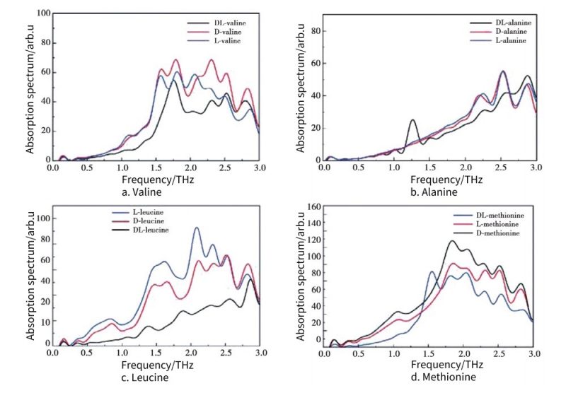
Fig. 7 THz absorption spectra of different amino acids
From the obtained spectrograms, it is found that although their structures are similar, there are still obvious differences in the absorption spectra.
It can be observed from Figure 7 that the peaks of the absorption spectra of different amino acids are different, the overall changes of the absorption waves are also different, and the corresponding energies are also different.
2.1.2 DNA
With the continuous development of terahertz spectroscopy in recent years, this technology can be used to detect the energy of different molecules, including DNA. Since DNA is a biological macromolecule, its biological energy is in the terahertz band, so it can be directly detected by terahertz.
This technology is currently widely used in the field of DNA. Since different terahertz bands have different absorption spectra for DNA molecules, Figure 8 is mainly for the terahertz absorption spectra of 2′-deoxycytidine nucleotide and 5-methylcytidine molecules.
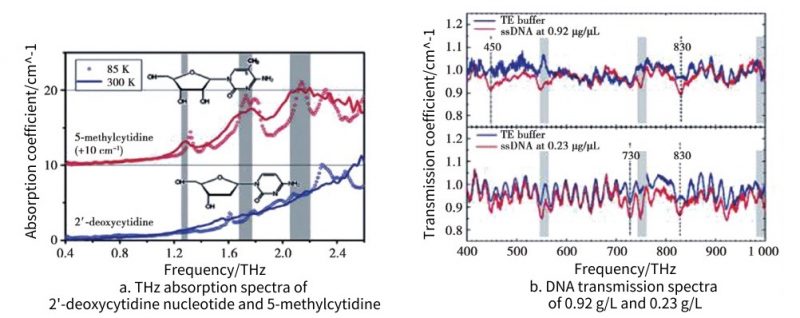
Fig. 8 THz spectra of different DNA
Zhang et al. studied a new terahertz spectroscopy technique, which can be used to obtain the transmission spectra of DNA in solution at different concentrations, it can be clearly observed from Fig. 8 that the transmission fluctuation range of the DNA at the concentration of 0.23 and 0.92 μg is different, and the transmission spectrum of the DNA at the concentration of 0.92 μg is relatively concentrated.
2.1.3 Chinese medicinal materials
Terahertz technology can not only be applied to biomacromolecules such as amino acids, but also has a wide range of applications in traditional Chinese medicine.
At present, this technology is mainly used in drug identification and quality control in the field of traditional Chinese medicine. For example, natural bezoar and artificial bezoar, different origins, ingredients and varieties of real and fake rhubarb, some researchers have tested the toxic components contained in Huangpu auramine at the level of quality control. Figure 9 shows the absorption spectra of Angelica sinensis, Poria cocos, Citrus aurantium and Radix isatidis obtained by terahertz time-domain spectroscopy.
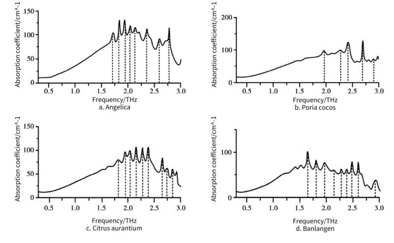
Fig. 9 Absorption spectrum of medicinal herbs
The results show that the optical properties of the three herbs are significantly different in the 0.2-2.2 THz band. Researchers at Peking University used support vector machine-based terahertz time-domain spectroscopy combined with principal component analysis to identify the medicinal herbs Chuanxiong and Fuxiong in Sichuan and other places, and the classification accuracy rate was 99.9%.
Researchers from China Agricultural University studied 4 kinds of tulips, golden turmeric, green turmeric, warm turmeric and osmanthus turmeric. The research results show that Chinese medicinal materials with similar properties can be identified using terahertz technology. Researchers at Tsinghua University used terahertz time-domain spectroscopy to detect gastrodin from two origins. The experimental data showed that the terahertz spectra of gastrodin extracted from two different origins of Gastrodia elata were different.
Researchers at Peking University used this technology to classify and identify grass saffron and saffron, with an accuracy of 100%, and for the identification of artificial and natural bezoar, the accuracy was as high as 100% and 90%, respectively.
2.2 Application of terahertz imaging technology
2.2.1 Epidermal tissue
Terahertz waves have high sensitivity and accuracy in the detection of skin moisture content. Detect the water content in the skin through terahertz waves, so as to understand the state of the skin, including skin dryness, oversaturation, skin inflammation and other problems.
Generally speaking, the water content distribution of pathological skin, such as skin inflammation, skin allergy or burn, is obviously different from that of normal skin. The higher the water content in skin and other tissues, the more gradual changes in tissue structure occur as the density of cells, proteins, and peptides changes. Based on these favorable factors, terahertz spectroscopy lays the foundation for imaging diagnosis of skin burns.
Arbab and other researchers from the University of Washington in the United States carried out terahertz time-domain spectroscopy experiments on mouse skin with different degrees of burns. And generate imaging images of mouse skin cells with burn degrees of 1, 2, and 3 degrees respectively, and compare them, which cover the absorption spectrum and reflection spectrum. Experiments have found that for different degrees of burns, at 0.5-0.7 THz, the terahertz absorption spectrum of mouse epidermal burn tissue is greater, and shows a higher reflectivity than terahertz waves.
In the frequency range of this band, 1 degree and 2 degrees have lower reflectivity and less absorption, Compared with skin tissue without burns, the reflectivity of 3rd degree burns is as high as 30%, which may be due to the higher water content of the cells and tissues at the burn site, forming edema.
In the time-dependent study on the degree of burn tissue, it was found that the terahertz reflectivity can reflect the degree of burn on the third day after burn, Correlations with burn severity were weaker in previous assays, suggesting that spectroscopic data confirmed by burn severity ratings were not obtained by immediately measuring skin wounds.
In addition, in the terahertz research of epidermal tissue, some researchers have conducted experiments on skin hair follicles, sweat gland structures, proteins in skin cells, DNA polysaccharides and amino acids, etc. And according to the approximate theory of Brugman’s dielectric constant and the double Debye dielectric relaxation model to explain the change of the dielectric function of different skin layers, the terahertz signal reflected by the skin surface was simulated.
2.2.2 Tumor research
In terms of medical applications, the advantages of terahertz waves are more significant, such as low penetrability to the human body and strong non-ionization, which lay the foundation for the non-destructive and rapid diagnosis of human tissues by terahertz technology . Cheon et al found that the use of terahertz microscopic imaging technology to obtain the fingerprint of superficial soft tissue tumors plays an important role in the early detection of cancer.
In 2018, Tyler et al. used terahertz imaging technology to identify cancer tissues that could assist in correcting pathological findings. Yeo et al. used terahertz imaging technology to study human lung and intestinal malignant tumors, and obtained the results that it can assist endoscopy in further clinical practice. At present, imaging is an important detection method for diagnosing early cancers, but traditional medical diagnostic techniques are more harmful to the human body.
Use this method to perform terahertz spectral imaging on normal liver tissue and tumor tissue, as shown in Figure 10, in which the white part is tumor tissue, and the black part is normal tissue, so that the terahertz imaging of normal tissue and tumor tissue has a more obvious effect.
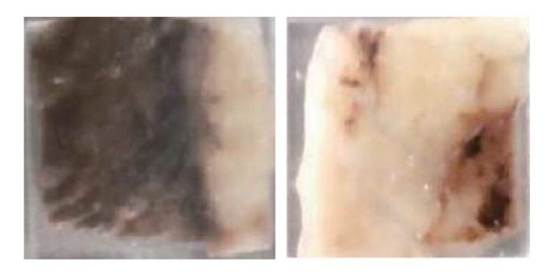
Fig. 10 Normal liver tissue and tumor tissue
The photoconductive THz-TDS system was used to detect the THz spectrum of normal tissue and tumor tissue. The results showed that the observed tissue sections were clearer when using the THz time-domain spectroscopy technique, as shown in Figure 11.
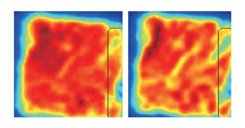
Fig. 11 Normal tissue and tumor tissue under terahertz imaging
Compared with normal tissue imaging, the tumor tissue imaging part is represented by a black rectangular area in Figure 11. From the color point of view, the color of normal tissue is orange-red, but according to the imaging results, the transmittance of tumor tissue is higher than that of normal tissue.
Terahertz imaging of liver tumor tissue is shown in Figure 12, Figure 12a-Figure 12c.
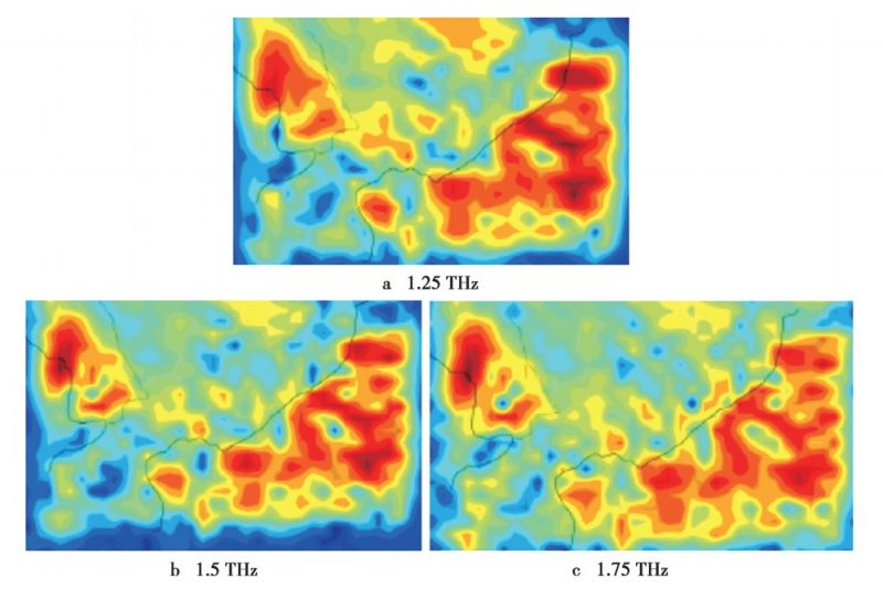
Fig. 12 Terahertz imaging of liver tumor tissue
They are the imaging results of liver tumor samples detected at 1.25, 1.5 and 1.75 THz respectively. From the images at 1.25 THz in Figure 12, it can be seen that the contrast of the imaging is poor at low frequencies.
As the frequency increases, the edge scattering of the imaged part gradually weakens, and the sharpness and contrast of the image are also improved. Therefore, within a reasonable imaging range, choosing the high-frequency position as much as possible is the key to obtaining high-quality imaging.
2.2.3 Bone Density Study
French M. Bessou et al. used THz-CT technology to realize bone terahertz imaging research.
Based on two-dimensional terahertz imaging, the team developed a new three-dimensional terahertz-band computed tomography method for imaging dry human bones, Under the statistics of countless experimental data, various parameters related to the refractive index and absorption rate of the terahertz spectrum are obtained, which have great practical significance in bone density analysis, which cannot be obtained in traditional X-ray technology detection . Human skeleton imaging is shown in Figure 13.
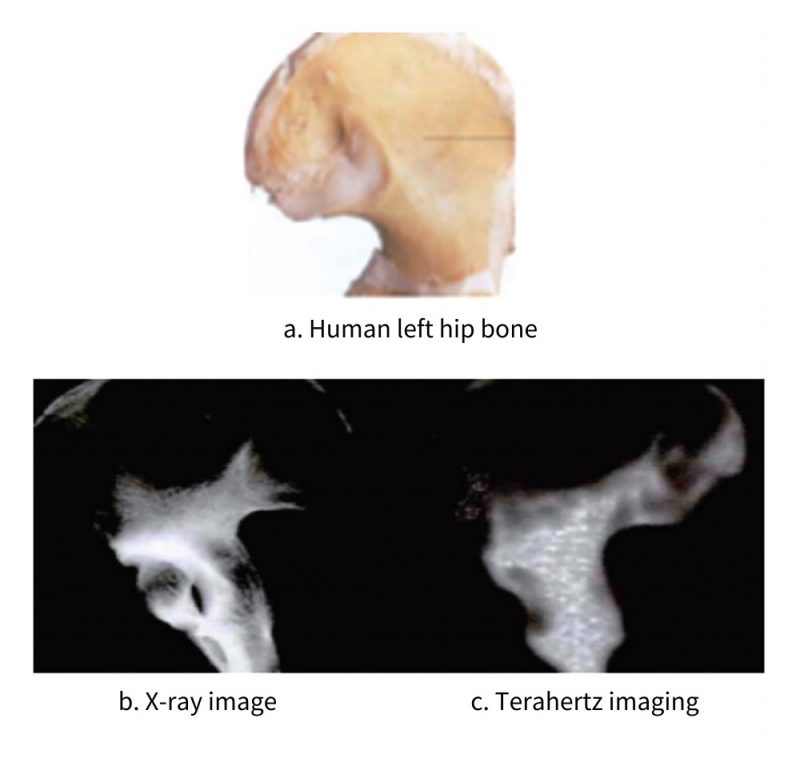
Fig. 13 Skeletal imaging of human body
Figure 13b and Figure 13c are X-ray imaging and terahertz imaging of the left hip bone respectively. It can be seen that the X-ray of the hip bone is highly transmissive, and the lower part of the hip bone appears to be opaque.
In the terahertz imaging of Figure 13c, it can be clearly seen that the hip bone wing contains more spongy tissue and the lower part of the hip bone contains more compact bone. This experiment demonstrates that terahertz imaging is capable of distinguishing between compact and cancellous bone.
2.2.4 The influence of terahertz radiation heat effect on biological cells
The influence of terahertz thermal radiation on biological tissue cells has also attracted extensive attention from researchers from all over the world.
G.C. Ferreira of the University of Liverpool in the UK studied the effects of different power terahertz radiation on human cells and tissues. Researchers at Harvard Medical School and Los Alamos National Laboratory have proposed that terahertz detection technology is likely to conduct individual contact with cells in an unmanned state, only through machines.
But at present this idea is only based on imagination and has not yet been proven. It can be seen that the technology in the field of terahertz is gradually applied to various researches on cells, and in the near future, significant results will be achieved.
3 Existing problems and future development direction
This paper mainly studies the two technologies of terahertz time-domain spectroscopy and terahertz imaging. Based on different detection methods for different samples, terahertz time-domain spectroscopy can be divided into two categories, namely transmission and reflection systems.
Terahertz imaging is divided into two types: pulse imaging and continuous wave imaging. Based on these two techniques, studies are mainly carried out on epidermal tissues, tumors, cells, etc.
It can be obtained that in the range of 0.5-0.7 THz, the terahertz absorption spectrum of epidermal burn tissue is larger and shows higher reflectivity than terahertz waves. In traditional Chinese medicine, terahertz time-domain spectroscopy is used to identify different herbal medicines.
However, after a large number of experimental operations and discussions among researchers, terahertz technology has developed rapidly, especially in the medical field.
However, there are still many problems to be explored and studied:
- Medical diagnosis is limited to specific research, and there is a lack of universal research on most cases. Due to the limitation of the current terahertz tuning range, low-frequency bands (<4THz) are mostly used for medical research.
Therefore, based on this phenomenon, it is also necessary to conduct research on high-frequency terahertz waves in the fields of tumors and tissues, which can be used for spectrum analysis and imaging; - The degree of water absorption.
Water absorbs terahertz waves very strongly, and the solution environment of biomolecules causes great noise, resulting in poor sensitivity,
Even the effective terahertz response signal cannot be collected, and the terahertz response spectrum of biological macromolecules cannot be detected in the state of keeping the stock solution of water.
Therefore, a breakthrough is needed in this respect; - In terms of medical diagnosis, terahertz detection may be used as an effective supplement to the existing imaging and pathology in the early diagnosis of tumors and the judgment of surgical margins. However, the application in this area is still in its infancy, and the research results are limited.
In response to these problems, in the future development stage of terahertz, the following goals are proposed:
- Increase the overall development of terahertz in the medical field, increase the scope of application and popularize data;
- Improve the resolution of imaging to make imaging clearer in medicine;
- Combining imaging technology with the detection of specific proteins and nucleic acids to develop new detection instruments;
- Real-time non-invasive in vivo medical imaging and in vivo objective case detection.
4 Summary and Outlook
This article first introduces terahertz imaging and spectroscopy technology, and then summarizes the research of this technology in the fields of human skin tissue, tumor, bone, biological cells, and traditional Chinese medicine.
It laid the foundation for the application of terahertz technology in the medical field in the future. Judging from the current research results, terahertz technology has great application value in medical detection and diagnosis, and will become a very important detection method in the medical field.
However, according to the current research progress, terahertz spectroscopy and imaging technology still needs a long period of research before it can be truly applied in clinical practice. At present, with the vigorous development of terahertz devices, terahertz spectroscopy and imaging technologies have also developed rapidly.
Many research institutions at home and abroad have carried out research work on its applications in multiple fields such as spectroscopy and imaging, which has further promoted the development of the terahertz field.
Although my country’s development in this field started relatively late, with the continuous efforts of researchers, some results have been achieved, and there is still a gap, which requires the joint efforts of researchers at home and abroad in the future. I believe that terahertz spectral imaging technology will benefit all mankind